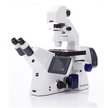
PHI Versaprobe II X-Ray Photoelectron Spectroscopy
Model: PHI Versaprobe II XPS
Function: The PHI VersaProbe II Scanning X-ray Microprobe system is a state-of-the-art Multi-technique instrument. The system provides X-ray Photoelectron Spectroscopy (XPS) wherein samples are irradiated with monochromatic X-rays, causing photoelectrons to be emitted from a surface of a sample. An electron energy analyzer then determines the binding energy of the photoelectrons which provides elemental, chemical state, surface, topographic, bulk chemical and interfacial analysis. The technique is compatible with a range of sample classes. The PHI VersaProbe II employs a unique high flux X-ray source that utilizes a focused electron beam scanned upon an Al anode for X-ray generation and a quartz crystal monochromator that focuses and scans the generated X-ray beam upon the sample surfaces to provide outstanding micro-spectroscopy analytical performance. It is designed for rapid, spatially resolved (micron scale) elemental and chemical analysis of surfaces. The PHI VersaProbe II combines a unique monochromatic X-ray probe with automated sample handling and analysis area identification tools to give true chemical state imaging and multi-point spectroscopy in an automated environment. For sample neutralization, the electron beam scans the anode surface at very high speed to reduce differential charging effects that cause data interpretation problems with non-conductive samples.The PHI VersaProbe II is also equipped with an argon sputtering gun for depth analysis of samples, a hot/cold stage for temperature dependent analyses, and an air-sensitive sample transfer vessel. The instrument is also equipped with Ultraviolet Photoelectron Spectroscopy (UPS) which provides information about the valence region of samples as well as the work function for materials.
Features: The PHI VersaProbe II Scanning X-ray Microprobe system is a state-of-the-art Multi-technique instrument. The system provides X-ray Photoelectron Spectroscopy (XPS) wherein samples are irradiated with monochromatic X-rays, causing photoelectrons to be emitted from a surface of a sample. An electron energy analyzer then determines the binding energy of the photoelectrons which provides elemental, chemical state, surface, topographic, bulk chemical and interfacial analysis.
Contact:
- Unit: Nanoscale Characterization Facility
- Campus:
- Bloomington
- Resource Type:
- Equipment
- Contact Name: Lyudmila Bronstein
- Contact Email: lybronst@indiana.edu
Return to Search

